A 10-foot microscope reveals big lessons about the tiniest threats to the human body
A 10-foot microscope reveals big lessons about the tiniest threats to the human body
There’s a technological revolution underway that’s making it faster and easier for scientists to see the molecules that undermine human health — and possibly fight the problem.
The “resolution revolution” involves cryo-electron microscopes, whose ever-improving detectors and software are producing three-dimensional images in unprecedented detail, aiding drugmakers.
They reveal detail so precise that biologist Andrew Ward, of Scripps Research in La Jolla, California, was able to spotlight the Achilles’ heel of several types of coronaviruses in images that he produced in 2016-17.
The weaknesses he called out? Spike proteins — the now-familiar elements that allow such viruses to infect cells.
This helped drugmakers to know exactly what to target when SARS-CoV-2 — the coronavirus that causes COVID-19 — emerged in late 2019.
Ward produced an even clearer snapshot of the proteins in 2020, further helping scientists create vaccines.
With additional help from him, effective vaccines were quickly produced.
“That was just the beginning,” said Ward. “Now, this technology is opening doors that help us understand the roots of diseases like cancer and neurodegeneration, including Alzheimer’s.”
“It routinely lets us see life’s tiniest machines — proteins, viruses and the atoms they combine — with breathtaking clarity,” he added.
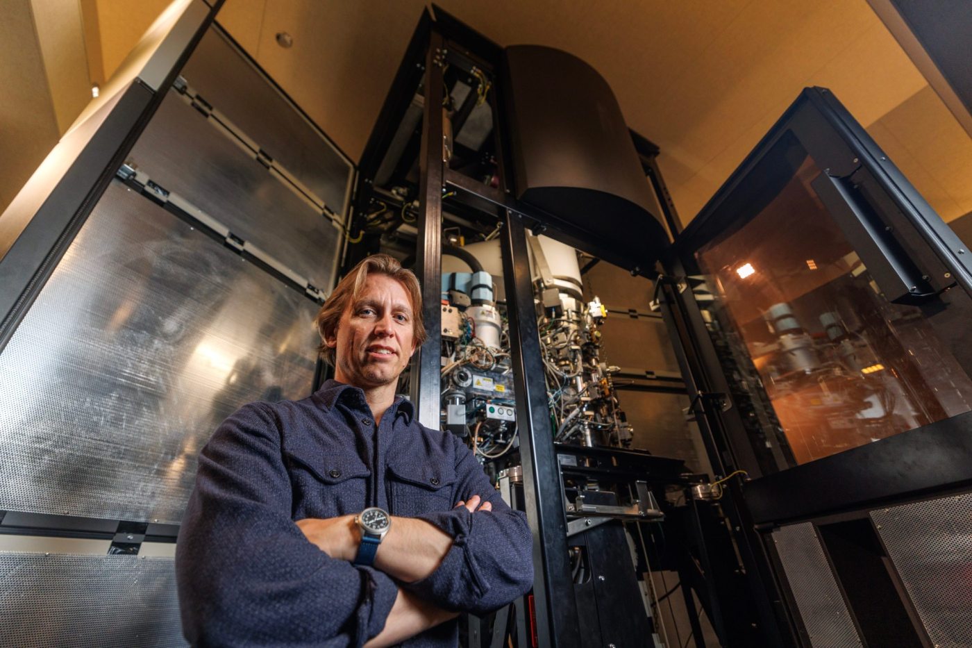
The research isn’t widely known to the public, partly because it’s hard to conceive of how any microscope — let alone one that’s 10 feet tall — can flash-freeze moving molecules, exposing their structure and purpose.
Ward offered a simple analogy to explain the matter.
“Imagine walking into a dark room,” he said. “You can roughly tell where the furniture is, and see shadowy outlines of a couch or a table. But once the lights are on, you can visualize color, texture, size and fine details.”
That’s what cryo-electron microscopes do — and with great speed.
Ward could only produce about 200 images per day when he was earning his doctorate at Scripps from 2003 to 2008, when he was using a far less powerful type of cryo-electron microscope. And he had access to it only a day or two each month.
Today, he can generate 1,500 images per second on Titan Krios, the largest and most powerful of Scripps’ seven cryo-electron microscopes.
If you were able to stack up the images he takes during a six-hour period, they would rise as high as Mount Everest, said Ward, who has been collaborating with institutes on SARS-CoV-2, Lassa, HIV, malaria and the H5N1 bird flu.

To the unfamiliar eye, the images look bizarre. Some resemble bumpy, frozen lava, others crinkly Christmas wreaths. Still others look like the knotty cords on old landline telephones.
But their importance is understood by scientists, especially those focused on preparing the world for whatever pandemic could come next.
It’s a bit of a fluke that Ward is a rising star at an institute that has helped develop more than 15 FDA-approved drugs and treatments, including Humira, which is used by people who suffer from arthritis.
He was interested in science growing up outside Boston — less so when he entered Duke University as a freshman.
Things quickly changed when he took a work-study job in a campus lab, where cell biologists Michael and Mary Reedy let him tinker. Before long, Ward was helping build the components of microscope cameras and detectors, and was dazzled by what they could do.
“I began to see molecules and atoms,” said Ward, now 46. “It kind of blows your mind to follow things at that resolution.”
Electron microscopes have existed since the 1930s, and they’ve played a vital role in revealing the structure of proteins and viruses and how they work. But the instruments didn’t start to enter their current golden age until roughly 2001, the year Ward arrived at Scripps as a lab technician.
The advancements have come especially quickly over the past decade, starkly improving image resolution and enabling scientists to see individual atoms. Software has also made it easier to see molecules interact with prospective drugs, helping determine which ones should go on to large-scale clinical trials.
The boom, locally and worldwide, didn’t go unnoticed. In 2017, three European scientists won the Nobel Prize in chemistry for helping transform cryo-electron microscopes into indispensable tools to explore the life sciences.
Ward says he’s happy to be in the background. But he has emerged as a leader in the field — mostly through his use of Titan Krios.
“Big Daddy,” as Ward calls it, is highly sensitive. The towering microscope sits on stabilizers to prevent something as simple as a slammed door from producing vibrations that could mess up image-taking. It operates in silence for the same reason.

In plain terms, the microscope freezes biological samples, then hits them with electron beams that create images.
“Once you see the arrangement of atoms, the connectivity of molecules, you can become an engineer,” Ward said. “You can move things around and manipulate the building blocks of life to make new therapeutics and vaccines that have much higher likelihood of success compared to engineering without blueprints.
“We’ve sped up the process of choosing which one should be a go, or no-go, for clinical trials,” Ward said.
That doesn’t mean scientists are close to flooding the market with new means of prevention.
“The potential vaccines will collectively have to go through five to seven years of trials in humans,” he added. “But we are no longer shooting in the dark or relying on empiricism.
“We can now shine a light — or rather a very powerful electron beam — on the science driving vaccine research.”


With Beyoncé's Grammy Wins, Black Women in Country Are Finally Getting Their Due
February 17, 2025


Comments 0